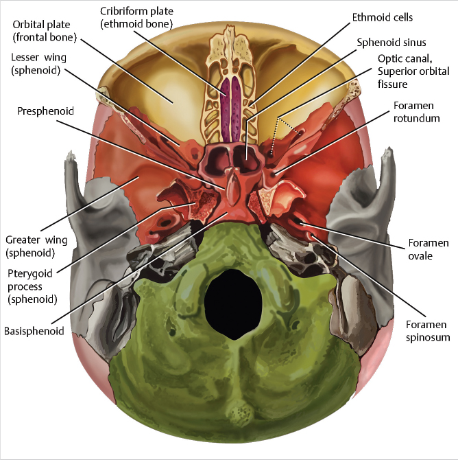
Cranial cavity is a space within the skull that houses and protects the brain.
The cranial cavity is a part of the human skull that houses and protects the brain. It is a hollow space within the skull, enclosed by the cranial bones. The cranial cavity is lined with three layers of protective membranes called meninges, which provide additional cushioning and support for the brain.
The cranial cavity is bounded by the frontal, parietal, temporal, and occipital bones of the skull. These bones form a rigid structure that safeguards the brain from external impacts and injuries. The cranial bones are joined together by sutures, which allow for minimal movement and provide stability.
The brain takes up the majority of the room inside the cranial cavity. The brain is the hub of the nervous system and is in charge of directing and coordinating all body processes. It is made up of several parts, such as the brainstem, cerebellum, and cerebrum, each of which performs a certain purpose.
Various cranial nerves, blood arteries, and cerebrospinal fluid (CSF) are among the other components found in the cranial cavity. While the CSF circulates around the brain and spinal cord, providing extra protection and preserving a stable environment, the blood arteries deliver oxygen and nutrients to the brain.
It is also known as the intracranial cavity. It is made up of three distinct depressions i.e.
- Middle cranial fossa
- Anterior cranial fossa
- Posterior cranial fossa

To simplify the understanding of the divisions of the cranial cavity, you can think of it as having three major compartments or regions:
- Anterior Cranial Fossa: This is the front part of the cranial cavity, located at the base of the frontal bone. It accommodates the frontal lobes of the brain, which are responsible for various cognitive functions, personality, and voluntary movement.
- Middle Cranial Fossa: This is the central part of the cranial cavity, situated behind the anterior cranial fossa. It houses structures like the temporal lobes, the pituitary gland, and the optic chiasm. The temporal lobes are involved in processes such as memory, hearing, and language.
- Posterior Cranial Fossa: This is the rear part of the cranial cavity, located at the base of the skull. It contains the cerebellum, brainstem, and part of the occipital lobes. The cerebellum is responsible for motor coordination and balance, while the brainstem regulates essential functions like breathing, heart rate, and consciousness.
The arrangement of the cranial cavity and the primary components it houses may be more easily understood by segmenting it into these three areas. Keep in mind that the many divisions of the cranial cavity support and protect the various brain regions, each of which has a unique set of functions.
(a) Anterior cranial fossa.
It is the most shallow and superior of the three cranial fossae and lies superiorly over the nasal and orbital cavities.
Houses the projecting frontal lobes of the brain.

Boundaries.
Anterior cranial fossa is bounded as follows.
- Anterior and laterally
Inner surface of the frontal bone.
- Posteriorly and medially
Limbus of the sphenoid bone- Limbus is a bony ridge that forms the anterior border of prechiasmatic sulcus.
- Posteriorly and laterally
Lesser wings of the sphenoid bone.
- Floor
Consists of the frontal bone, ethmoid bone and anterior aspects of the body and lesser wings of sphenoid bone.
Contents.
Anterior cranial fossa contains the following parts of the brain.
-Frontal lobe of the cerebral cortex
-Olfactory bulb
-Olfactory tract
-Orbital gyri
Crista galli is an upward-projecting bone that is located in the middle of the ethmoid bone. It serves as yet another attachment place for the falx cerebri.
Crista galli has a cribriform plate on either side. In addition to having multiple foramina that transport nerves and arteries, it supports the olfactory bulb.
Lesser wings protrude from the anterior sphenoid bone’s central body. The tentorium cerebelli is attached to the smaller wings by the clinoid processes, which have rounded ends.
Frontal crest, a bodily ridge, denotes the frontal bone in the midline.
The frontal crest protrudes upward and serves as the falx cerebri’s attachment point.
Foramina.
Ethmoid bone in particular contains the main foramina of the anterior cranial fossa.
(i) Cribriform foramina.
Are openings in the cribriform plate of the ethmoid bone, which connect the anterior cranial fossa with nasal cavity and transmit the olfactory nerves.
(ii) Anterior ethmoid foramen.
Connects anterior cranial fossa with each orbit.
Transmits the anterior ethmoidal artery, nerve and vein.
(iii) Posterior ethmoidal foramen
Transmits the posterior ethmoidal artery, nerve and vein.
(b) Middle cranial fossa.
Boundaries.
Formed by sphenoid bones and temporal bones.
(i) Anterior
Lesser wings of the sphenoid bone
(ii) medially
Limbus of the sphenoid bone
(iii) Posteriorly and medially
Dorsum sellae of the sphenoid bone-It’s a large superior projection of the bone that arises from sphenoidal body.
(iv) Floor – greater wings of sphenoid,
-squamous and petrous parts of temporal bone.

Contents.
Middle cranial fossa consists of a central portion, which contains the pituitary gland and two lateral portions which accommodate the temporal lobes of the brain.
-Central part of middle cranial fossa formed by sphenoid bone contains Sella turcica.
-Sella turcica acts to hold and support the pituitary gland.
-It consists of the three parts
(a) Tuberculum sellae -A vertical elevation of bone that forms anterior wall of the Sella turcica and the posterior aspect of the chiasmatic sulcus.
(b) Hypophyseal fossa/Pituitary fossa- Middle of Sella turcica. It holds the pituitary gland.
(c)Dorsum sellae- Forms posterior wall separates middle cranial fossa from posterior cranial fossa.
The depressed lateral parts are formed by greater wings of the sphenoid, squamous and petrous parts of temporal bones.
They support temporal lobes of the brain.
Foramina.
(a)Foramina of the sphenoid bone.
(I)Optic Canals-Situated anteriorly in the middle cranial fossa.
Transmits: Optic nerves CN II
Ophthalmic arteries.
(ii) Superior orbital fissure-Opens anteriorly into the orbit.
Transmits four cranial nerves:
- Oculomotor CN III
- Trochlear CN IV.
- Ophthalmic CN V
Abducens CN VI and
Ophthalmic veins
Sympathetic nerve fibers.
(iii)Foramen Rotundum – A paired opening serving as a connection between middle cranial fossa and pterygopalatine fossa.
Transmits: Maxillary nerve CN V2
(iv) Foramen Ovale-Paired opening connecting middle cranial fossa with external surface of cranial base and the infratemporal fossa.
Transmits: Mandibular nerve CN V3
Lesser petrosal nerve
Accessory meningeal branch of maxillary artery
Emissary vein
(v) Foramen Spinosum (paired) connects middle cranial fossa with infratemporal fossa.
Transmits: Middle meningeal artery and vein.
Meningeal branch of mandibular nerve CN V3
(b) Foramina of the temporal bone.
(i) Carotid canal-connects middle cranial fossa to external cranial base.
Carries: Internal carotid artery
Deep petrosal nerve also passes through this canal.
(iii)Hiatus of greater petrosal nerve
Transmits: greater petrosal nerve (branch of facial nerve)
Petrosal branch of middle meningeal artery.
(iv) Hiatus of the lesser petrosal nerve
Transmits: Lesser petrosal nerve -branch of the glossopharyngeal nerve
(v) Foramen Lacerum – found at the junction of sphenoid, temporal and occipital bone. Filled with cartilage, which is pierced only by small blood vessels.
(c) Posterior cranial fossa.
The posterior cranial fossa is essential for protecting and supporting vital structures involved in motor control, balance, and essential functions of the nervous system. Any abnormalities or injuries in this region can have significant implications for neurological health and well-being.
Boundaries.
(i)Anteriorly
Dorsum sellae of sphenoid bone
(ii) laterally
Superior border of petrous part of temporal bone.
(iii) Posteriorly
Internal surface of squamous part of occipital bone.
(iv) Floor
-mastoid part of temporal bone
-squamous, condylar and basilar parts of the occipital bone.
Contents.
Posterior cranial fossa contains:
- Cerebellum
- Brainstem
- Arteries
- Nerves
Foramina.
(a) Temporal bone.
(i) Internal acoustic meatus (paired)-connecting posterior cranial fossa and the facial canal.
Transmits: Facial nerve CN VII
Vestibulocochlear nerve CN VIII
Labyrinthine artery.
(b) Occipital bone
(I) Foramen magnum-largest opening in the skull connecting posterior cranial fossa with external cranial base.
Transmits: Lower end of medulla oblongata
- Vertebral artery
- Accessory nerve CN XI
- Anterior and posterior spinal arteries.
- Meninges
- Dural veins
(ii) Jugular foramina– Forms bilaterally between the occipital and temporal bones in the posterior cranial fossa and leads to the external cranial base.
Transmits: Inferior petrosal and sigmoid sinuses
- Glossopharyngeal CN I
- Vagus CN X
- Accessory nerve CN XI
(iii) Hypoglossal canal – leads to the external cranial base and carries the twelfth cranial nerve -hypoglossal nerve.
(iv) Cerebellar fossae– Found posteriorly to foramen Magnum. It’s a bilateral depression.
-Houses the cerebellum
-Divided medially by a ridge of bone, the internal occipital crest.
